Choledocholithiasis Ultrasound - Physical exam reveals tenderness to palpation in the right upper quadrant and negative murphy sign. Complete blood count (cbc) liver function tests; Tests that show the location of stones in the bile duct include the following: A test that could demonstrate choledocholithiasis is an mrcp (magnetic resonance cholangiopancreatography) or an ercp (endoscopic retrograde cholangiopancreatography). Pancreatic enzymes (amylase or lipase) You should also read this: Planned Parenthood Tb Test

The Role of Laparoscopic Ultrasonography in the Evaluation of Suspected - Magnetic resonance cholangiopancreatography (mrcp) percutaneous transhepatic cholangiogram (ptca) The presence of choledocholithiasis is confirmed by imaging studies such as transabdominal ultrasound (low sensitivity for choledocholithiasis in the distal common bile duct), contrast enhanced abdominal computed tomography (ct) scan, magnetic resonance cholangiopancreatography (mrcp), intraoperative cholangiogram. Eus is less invasive than ercp, and mrcp is noninvasive. Ct scanning is only 75% sensitive. You should also read this: Emission Test In Bridgeport Ct

Choledocholithiasis MEDizzy - Ct scanning is only 75% sensitive for choledocholithiasis and not the test of choice. Blockage and infection caused by stones in the biliary tract can be life threatening. However, ercp with sphincterotomy is most commonly employed with a high degree of success. A transabdominal ultrasound is the first test that should be ordered for the patient suspected of any biliary. You should also read this: Psa Test Reliability

Choledocholithiasis an Overview - To demonstrate choledocholithiasis, blood tests can be done to look for high levels of bilirubin and liver enzymes. Transabdominal ultrasound is insensitive for choledocholithiasis as opposed to its excellent performance in cholelithiasis. Endoscopic retrograde cholangiography (ercp) endoscopic ultrasound; Ct scanning is only 75% sensitive for choledocholithiasis and not the test of choice. Diagnosis of choledocholithiasis is not always straightforward and. You should also read this: Hillsborough County Driving Test

Choledocholithiasis Ultrasound - Tests that show the location of stones in the bile duct include the following: To demonstrate choledocholithiasis, blood tests can be done to look for high levels of bilirubin and liver enzymes. Physical exam reveals tenderness to palpation in the right upper quadrant and negative murphy sign. Pancreatic enzymes (amylase or lipase) The goal of treatment is to relieve the. You should also read this: Does Fsa Cover Pregnancy Tests

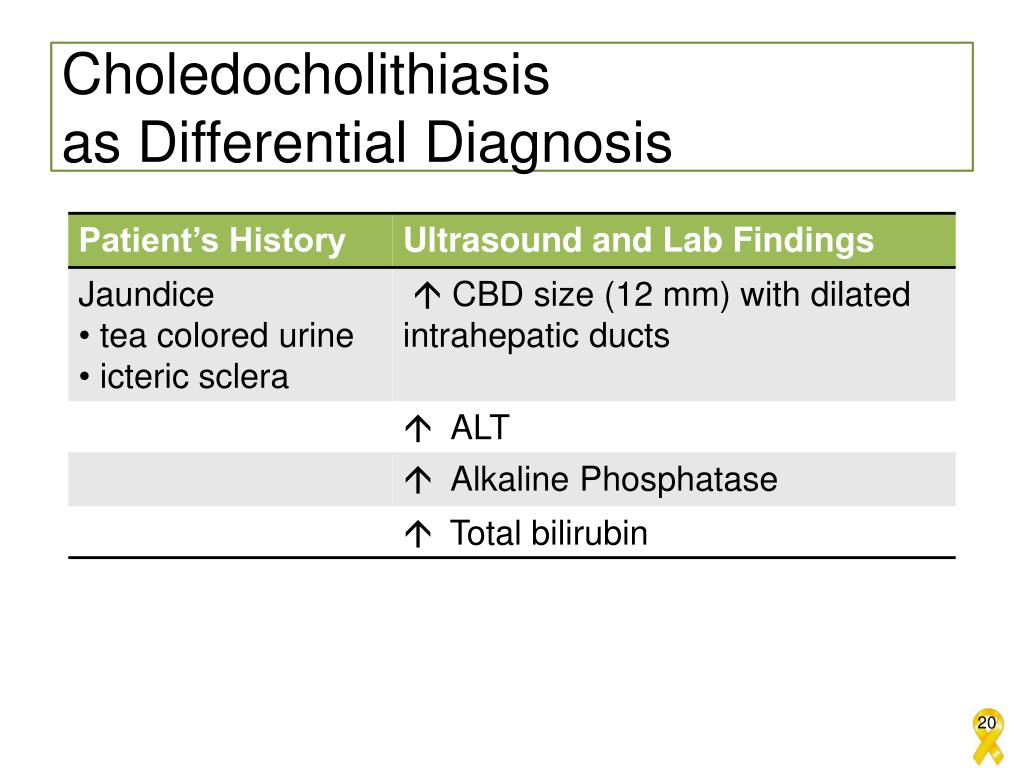
Clinical predictors of choledocholithiasis.CBD common bile duct, US - She is admitted and scheduled for an mrcp. Laboratory results show increased alkaline phosphatase and total bilirubin. A right upper quadrant ultrasound shows a dilated common bile duct, suggestive of choledocholithiasis. Endoscopic retrograde cholangiography (ercp) endoscopic ultrasound; The presence of choledocholithiasis is confirmed by imaging studies such as transabdominal ultrasound (low sensitivity for choledocholithiasis in the distal common bile duct),. You should also read this: Coolant Test Strips Autozone

PPT Choledocholithiasis PowerPoint Presentation, free download ID - For decades, endoscopic ascending retrograde cholangiopancreatography has been the golden diagnostic standard in cases of suspected choledocholithiasis. Confirmatory diagnosis of choledocholithiasis is made with advanced imaging, including magnetic resonance cholangiopancreatography and endoscopic retrograde cholangiopancreatography (ercp). Tests that show the location of stones in the bile duct include the following: Blockage and infection caused by stones in the biliary tract can. You should also read this: Dsp Orientation Test Answers

Choledocholithiasis Image - Laboratory results show increased alkaline phosphatase and total bilirubin. In most cases, an abdominal ultrasound will show a dilated common bile duct (more than 6. The method is associated with a relatively high rate of complications, including acute pancreatitis, the incidence of which is estimated to range between 0.74% and 1.86%. Physical exam reveals tenderness to palpation in the right. You should also read this: Website Automated Testing

Accuracy of MDCT in the Diagnosis of Choledocholithiasis AJR - Eus is less invasive than ercp, and mrcp is noninvasive. The presence of choledocholithiasis is confirmed by imaging studies such as transabdominal ultrasound (low sensitivity for choledocholithiasis in the distal common bile duct), contrast enhanced abdominal computed tomography (ct) scan, magnetic resonance cholangiopancreatography (mrcp), intraoperative cholangiogram. She is admitted and scheduled for an mrcp. In most cases, an abdominal ultrasound. You should also read this: Applause Testing Jobs

Choledocholithiasis Ultrasound - He or she may use one of the following imaging tests: To demonstrate choledocholithiasis, blood tests can be done to look for high levels of bilirubin and liver enzymes. Magnetic resonance cholangiopancreatography (mrcp) percutaneous transhepatic cholangiogram (ptca) The presence of choledocholithiasis is confirmed by imaging studies such as transabdominal ultrasound (low sensitivity for choledocholithiasis in the distal common bile duct),. You should also read this: Test De Las 7 Identidades