Stress testing and noninvasive coronary imaging What’s the best test - A narrowing ‘blockage” is normal at rest, and is present at stress, so that is a “reversible” defect. Background post placement is a common practice to reinforce weakened roots. The presence of a fixed perfusion abnormality was independently associated with an increased risk of death after adjustment for clinical and stress test data and the summed. Regional reductions in uptake. You should also read this: Cast Test For Lineman

The nuclear stress test using 99mc TC sestamibi scan showing the fixed - A normal spect stress demonstrates global, uniform isotope uptake at rest and then uniformly increased uptake at stress. Regional reductions in uptake during both stress. Ischemic left ventricular (lv) myocardium is detected as one or more perfusion defects arising during a stress test in a cardiac mri examination. A combination of technical and physiology. A combination of technical and physiology. You should also read this: Ibd Diagnostic Test

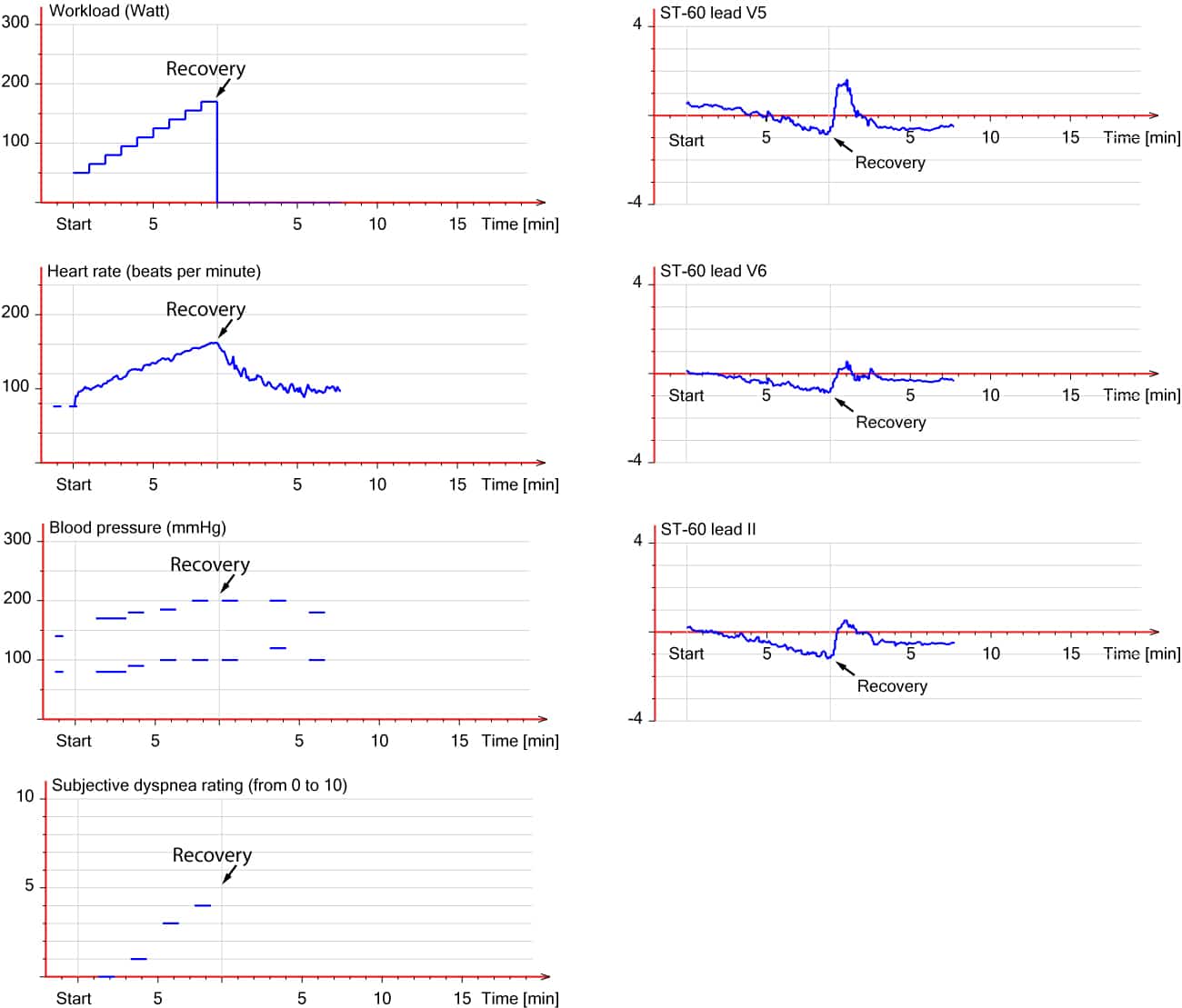
Evaluation of exercise stress test ECG, symptoms, blood pressure - Severe fixed defect most likely represents scarring or fibrosis from prior mi, but a mild. This brief review focuses on reasons why myocardial perfusion imaging (mpi) spect defects may appear “fixed” (rest vs stress). A narrowing ‘blockage” is normal at rest, and is present at stress, so that is a “reversible” defect. A combination of technical and physiology. Background post. You should also read this: B12 Test Tube Color

(A) Stress and rest myocardial perfusion SPECT short, vertical, and - Background post placement is a common practice to reinforce weakened roots. A normal spect stress demonstrates global, uniform isotope uptake at rest and then uniformly increased uptake at stress. Regional reductions in uptake during both stress. Ischemic left ventricular (lv) myocardium is detected as one or more perfusion defects arising during a stress test in a cardiac mri examination. A. You should also read this: Sprit Animal Test

99m Tcsestamibi images show a fixed defect in the inferior wall - This brief review focuses on reasons why myocardial perfusion imaging (mpi) spect defects may appear “fixed” (rest vs stress). Scarred myocardium from prior infarct will not take up tracer at all and is referred to as a fixed defect. stress mri is only performed with the infusion of adenosine and is a newer means of. A combination of technical and. You should also read this: Dime Test Kitchenaid

(A) Stress and rest myocardial perfusion SPECT short, vertical, and - A normal spect stress demonstrates global, uniform isotope uptake at rest and then uniformly increased uptake at stress. This brief review focuses on reasons why myocardial perfusion imaging (mpi) spect defects may appear “fixed” (rest vs stress). Scarred myocardium from prior infarct will not take up tracer at all and is referred to as a fixed defect. stress mri is. You should also read this: Gut Health Test With Microbiome Wipe

Nuclear stress test showing fixed perfusion defect in the inferior wall - This brief review focuses on reasons why myocardial perfusion imaging (mpi) spect defects may appear “fixed” (rest vs stress). A combination of technical and physiology. Regional reductions in uptake during both stress. An old heart attack leaves scar tissue, so this looks the same with rest and. Scarred myocardium from prior infarct will not take up tracer at all and. You should also read this: Testing Crossword Clue

Nuclear stress test showing fixed perfusion defect in the inferior wall - This brief review focuses on reasons why myocardial perfusion imaging (mpi) spect defects may appear “fixed” (rest vs stress). Scarred myocardium from prior infarct will not take up tracer at all and is referred to as a fixed defect. stress mri is only performed with the infusion of adenosine and is a newer means of. Regional reductions in uptake during. You should also read this: Iowa State Testing Centers

Stress testing and noninvasive coronary imaging What’s the best test - A narrowing ‘blockage” is normal at rest, and is present at stress, so that is a “reversible” defect. Ischemic left ventricular (lv) myocardium is detected as one or more perfusion defects arising during a stress test in a cardiac mri examination. This brief review focuses on reasons why myocardial perfusion imaging (mpi) spect defects may appear “fixed” (rest vs stress).. You should also read this: My Wellspan Test Results
82 Rb PET perfusion images show a fixed perfusion defect on both stress - Severe fixed defect most likely represents scarring or fibrosis from prior mi, but a mild. A fixed (irreversible defect) is characteristic of myocardial. An old heart attack leaves scar tissue, so this looks the same with rest and. Ischemic left ventricular (lv) myocardium is detected as one or more perfusion defects arising during a stress test in a cardiac mri. You should also read this: Mikaela Testa Thothub