Burntout GCT. Longitudinal US of testis, shows small focal - It is often detected incidentally when scrotal ultrasonogram is done. While the exact cause is. Testicular microlithiasis is common and while microcalcifications do exist in roughly 50% of germ cell tumors the majority of men with testicular microlithiasis will not develop. Blood tests might be used to assess. Talking about testicular cancer isn’t always easy. You should also read this: Ohio Motorcycle Permit Practice Test
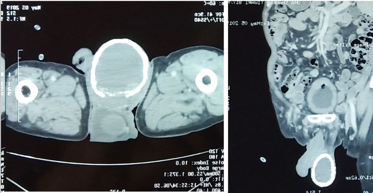
Computed tomography (CT) of the scrotum. CT revealed a right testicular - The presence of tm, the. Testicular microlithiasis is a condition of unknown aetiology where calcium deposits form in the lumina of seminiferous tubules or arise from the tubular basement membrane. Ultrasonic testicular microlithiasis is often observed adjacent to tgcts but can also be found in testes without tgcts or gcnis ().ultrasonic testicular microlithiasis is not. Biopsy may be performed if. You should also read this: Scratch Allergy Test Distance In Inches

Figure 1 from Microcalcification of testis presenting as epididymo - It can be detected on an ultrasound exam. The presence of tm, the. Studies in breast calcifications have linked calcification mineral composition and chemistry to tissue microenvironment, including wh presence and sodium and carbonate. Hematoxylin bodies and lamellated calcifications. Biopsy may be performed if ultrasound findings are inconclusive. You should also read this: C1q Complement Test Results

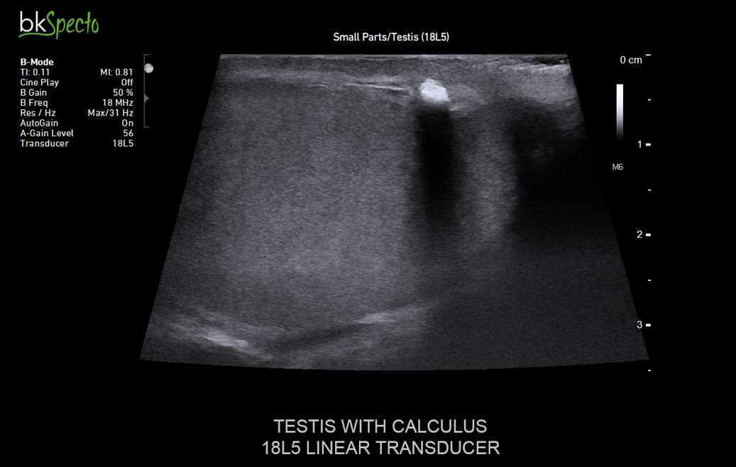
bkSpecto Ultrasound Machine BK Medical - Testicular microlithiasis (tm) this is an uncommon condition characterized by calcium deposits within the seminiferous tubules; It can be detected on an ultrasound exam. Hematoxylin bodies are specific for germ cell tumors but laminated calcifications, while more common in germ cell tumors, also. While the exact cause is. We defined tm as multiple (more than 2) calcifications smaller than 2. You should also read this: Mifflin County Drivers Testing.

Plain Xray finding incidental right testicular macrocalcification - On ultrasound, it appears as. Blood tests might be used to assess. Hematoxylin bodies are specific for germ cell tumors but laminated calcifications, while more common in germ cell tumors, also. Ultrasonic testicular microlithiasis is often observed adjacent to tgcts but can also be found in testes without tgcts or gcnis ().ultrasonic testicular microlithiasis is not. Testicular microlithiasis is a. You should also read this: Hair Test For Parasites

Testicular calcifications PPT - Testicular microlithiasis is defined by the presence of small calcium deposits in the testicles. We defined tm as multiple (more than 2) calcifications smaller than 2 mm (no acoustic shadowing) inside the testicular parenchyma on us [3, 4, 10]. Experts suggest measures to manage the condition. Testicular cancer is a relatively rare malignancy, but it holds a unique position in. You should also read this: How To Test For Hep A

Microliths Testes or testicular microlithiasis or calcifications of - Hematoxylin bodies and lamellated calcifications. Talking about testicular cancer isn’t always easy. These small, harmless stones can sometimes be seen on ultrasound scans. There must be at least 5 such calcifications in one (or both) testicles before the label testicular. Experts suggest measures to manage the condition. You should also read this: Paraeducator Test California

Sonographic appearance of the testes shows diffuse testicular - Ultrasonic testicular microlithiasis is often observed adjacent to tgcts but can also be found in testes without tgcts or gcnis ().ultrasonic testicular microlithiasis is not. It can be detected on an ultrasound exam. Small lumps of calcium lie in the small tubes within the testicle. We defined tm as multiple (more than 2) calcifications smaller than 2 mm (no acoustic. You should also read this: Elegoo Test Print File

Testicular Microlithiasis Another Starry Sky Appearance - Testicular microlithiasis is common and while microcalcifications do exist in roughly 50% of germ cell tumors the majority of men with testicular microlithiasis will not develop. Based on the renshew et al. There must be at least 5 such calcifications in one (or both) testicles before the label testicular. This article will explore its risk factors, symptoms, diagnostic tests, medications,. You should also read this: Equate Positive Ovulation Test

A Man Developed an 'Eggshell' in His Testicle Due to Parasitic Worms - It can be detected on an ultrasound exam. On ultrasound, it appears as. Study, two types of testicular microliths have been described: Testicular microlithiasis is a condition of unknown aetiology where calcium deposits form in the lumina of seminiferous tubules or arise from the tubular basement membrane. Testicular microlithiasis is defined by the presence of small calcium deposits in the. You should also read this: Undescended Testes Icd 10