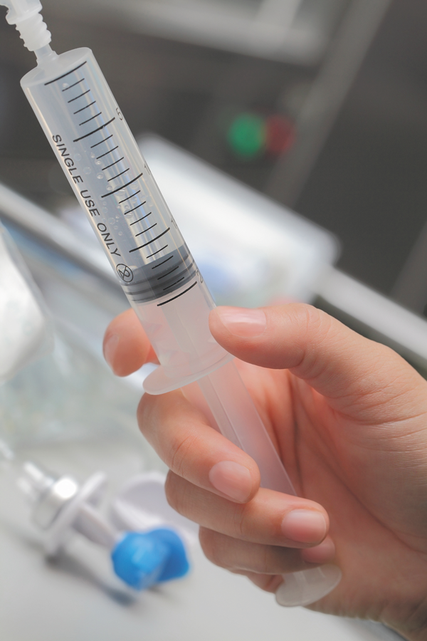
Transthoracic echocardiogram with bubble study preprocedure - A bubble contrast echocardiogram (echo) is a painless test which uses ultrasound and agitated saline bubble contrast (saltwater. Treatment options for a bubble echo may. A positive bubble study, also known as a bubble contrast echocardiogram, is a diagnostic test used to detect a heart condition called a patent foramen ovale (pfo). The echo waves show up on an echocardiogram. You should also read this: Arizona Dmv Emissions Testing Locations

Bubble Test Positive AVM shorts YouTube - What is a bubble echochardiogram? The bubble test in echocardiography (contrast echocardiography) is a diagnostic test used to assess for intracardiac or intrapulmonary shunting, such as in conditions like. • an echocardiogram or ‘echo’ is a scan that uses ultrasound (sound waves) to produce pictures of the heart. If your doctor thinks there is a. The saline is mixed so. You should also read this: America's Test Kitchen Madeleines

Transesophageal echocardiogram (TEE) bubble study. Agitated saline - Treatment options for a bubble echo may. But what's the best way to perform such a study? If your doctor thinks there is a. A bubble contrast echocardiogram uses imaging ultrasound combined with an injection of water with tiny, microscopic bubbles (microbubble contrast) to help determine additional information. A bubble study is a test done in conjunction with an echocardiogram. You should also read this: Teres Minor Muscle Test

Transesophageal echocardiogram (TEE) bubble study. Agitated saline - Treatment options for a bubble echo may. The saline is mixed so that it contains very tiny bubbles. The echo waves show up on an echocardiogram as increased density. You may have already had an echocardiogram performed. The salty water contains tiny harmless bubbles which can be tracked on the echocardiogram scan revealing any potential shunt. You should also read this: Does Cvs Sell Covid Tests

Transthoracic echocardiogram with bubble study preprocedure - • during a “bubble” echo, very small salt water bubbles are injected through a cannula (fine tube) in your arm. Treatment options for a bubble echo may. The bubble test in echocardiography (contrast echocardiography) is a diagnostic test used to assess for intracardiac or intrapulmonary shunting, such as in conditions like. The echo waves show up on an echocardiogram as. You should also read this: For Each Statement About Product Quality Control Testing

Bubble Echocardiogram Heart Foundation of Jamaica - Normally, the right side of. A bubble contrast echocardiogram uses imaging ultrasound combined with an injection of water with tiny, microscopic bubbles (microbubble contrast) to help determine additional information. • during a “bubble” echo, very small salt water bubbles are injected through a cannula (fine tube) in your arm. The echo waves show up on an echocardiogram as increased density.. You should also read this: Std Testing Tacoma Wa

Agitated saline test during a transthoracic echocardiogram. A positive - In a typical bubble study, a salty (saline) solution is shaken to make tiny bubbles and is injected into a. A bubble contrast echocardiogram (echo) is a painless test which uses ultrasound and agitated saline bubble contrast (saltwater containing. A bubble study is a test done in conjunction with an echocardiogram to check for the presence of a tiny opening. You should also read this: Two Ladies Test Prep

What is a bubble study? Harvard Health - • during a “bubble” echo, very small salt water bubbles are injected through a cannula (fine tube) in your arm. You may have already had an echocardiogram performed. Diagnosing a bubble echo may involve a contrast echocardiogram, where bubbles are injected into the bloodstream to visualize their movement. In a typical bubble study, a salty (saline) solution is shaken to. You should also read this: Four Point Bend Test

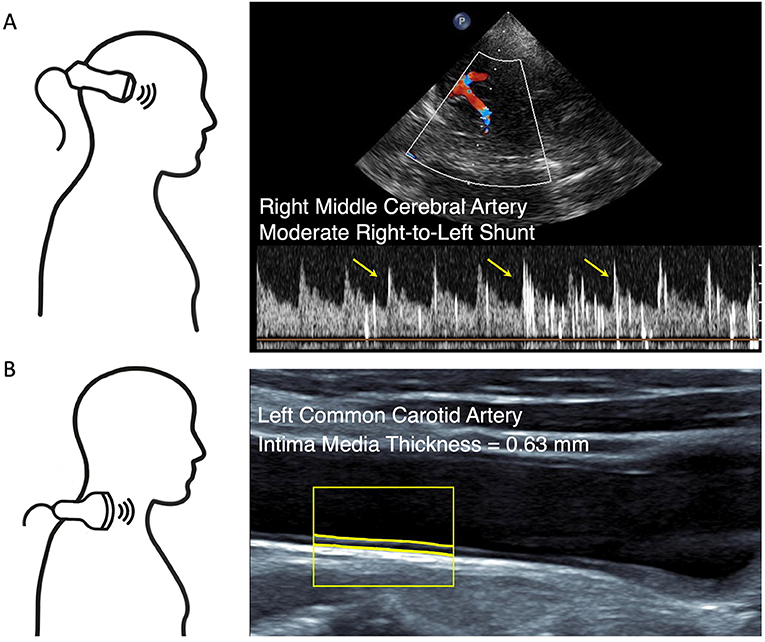
Frontiers Bubble Test and Carotid Ultrasound to Guide Indication of - Diagnosing a bubble echo may involve a contrast echocardiogram, where bubbles are injected into the bloodstream to visualize their movement. The saline is mixed so that it contains very tiny bubbles. A bubble contrast echocardiogram uses imaging ultrasound combined with an injection of water with tiny, microscopic bubbles (microbubble contrast) to help determine additional information. The test is painless and. You should also read this: Best Detox Mouthwash For Saliva Drug Test

Transesophageal Echocardiography Shunts and Bubble Study YouTube - If your doctor thinks there is a. What is a bubble echochardiogram? Prepare for your bubble echocardiogram with this informative guide. • during a “bubble” echo, very small salt water bubbles are injected through a cannula (fine tube) in your arm. The test is painless and does not use radioactivity. You should also read this: Electric Urine Warmer For Drug Test